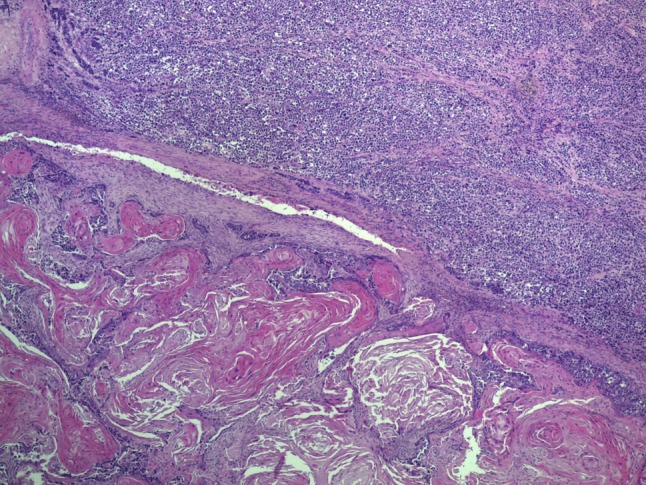
Fig. 2.

Low power view showing that the tumor consisted of two sharply separated components. The neuroendocrine carcinoma (top) consisted of solid sheets of poorly differentiated round to oval cells, while the squamous cell carcinoma component consisted of islands of atypical keratinizing epithelium (bottom)
