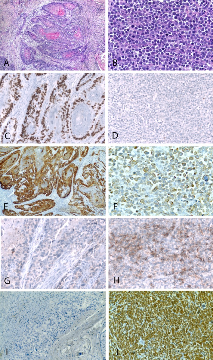
Fig. 3.
The squamous cell carcinoma component was characterized by a proliferation of invasive irregular solid tumor islands with evidence of keratinization (a). The neuroendocrine carcinoma consisted of a diffuse proliferation of poorly differentiated intermediate to large cells (b). Immunohistochemically, the squamous component was positive for p63 (c) while the neuroendocrine component was negative (d). Cytokeratin 5/6 (e) was expressed only in the squamous component, while the neuroendocrine component was positive for AE1/AE3 (f). The neuroendocrine carcinoma showed positivity for CD56 and chromogranin (h, j), while these markers were negative in the squamous component (g, i)

