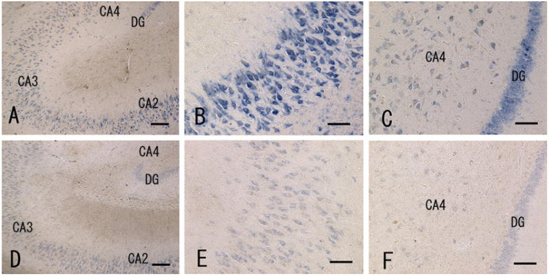
Fig. 2.

In situ hybridization of α1-chimaerin mRNA in the hippocampus of a control subject (A–C) and AD case (D–F). (A) and (D): at low magnification, positive signals were mainly visible in the pyramidal layers of the cornu ammonis (CA) and granular cell layer of the dentate gyrus (DG) in both control subjects and AD cases. (B) and (E): high magnification of the pyramidal layer of the CA2 region. (C) and (F): high magnification of the granular cell layer of the dentate gyrus. Signal intensity is reduced in the AD case (D–F) relative to that in controls (A–C). Scale bar = 200 μm in (A) and (D); 100 μm in (B, C, E and F).
