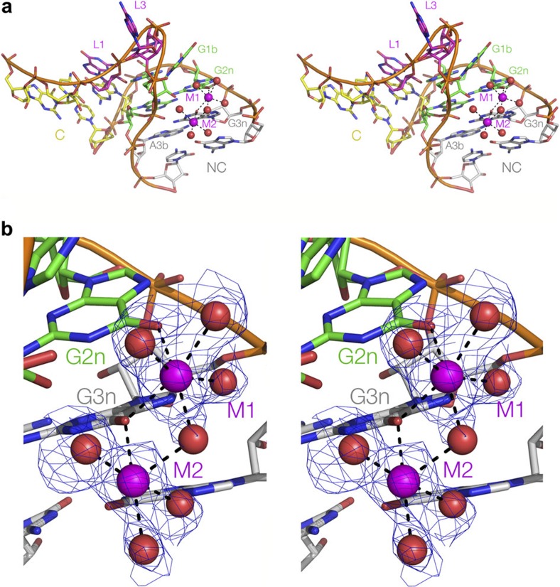
Figure 3. A structural basis for the differential ability of k-turn sequences to undergo ion-induced folding.
(a,b) Crystal structure of Kt-7 reveals two Mg2+ ions bound in the major groove of the NC helix. The structure is shown as parallel eye stereographic images. (a) An overall view of the k-turn, with bound ions. (b) Closer view of the bound ions. The electron density for the metal and directly bound water is taken from the Fo−Fc omit map contoured at 2σ. Further maps are shown in Supplementary Figure 5. Both ions have exchanged inner-sphere water ligands to bond to guanine O6 atoms; M1 is directly bonded to O6 of both G2n and G3n, while the latter is directly bonded to both ions.

