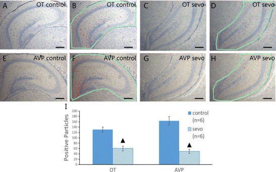
Fig. 4.

Immunohistochemical staining of OT and AVP in the dorsal hippocampus. a, b OT immunohistochemical staining results and the corresponding marking illustrations of positive particles in the air-treated group. c, d OT immunohistochemical staining results and the corresponding marking illustrations of positive particles in the sevoflurane-treated group. e, f AVP immunohistochemical staining results and the corresponding marking illustrations of positive particles in the air-treated group; g, h AVP immunohistochemical staining results and the corresponding marking illustrations of positive particles in the sevoflurane-treated group. (×100; scale bar, 200 μm, ▲ P < 0.001 compared between groups)
