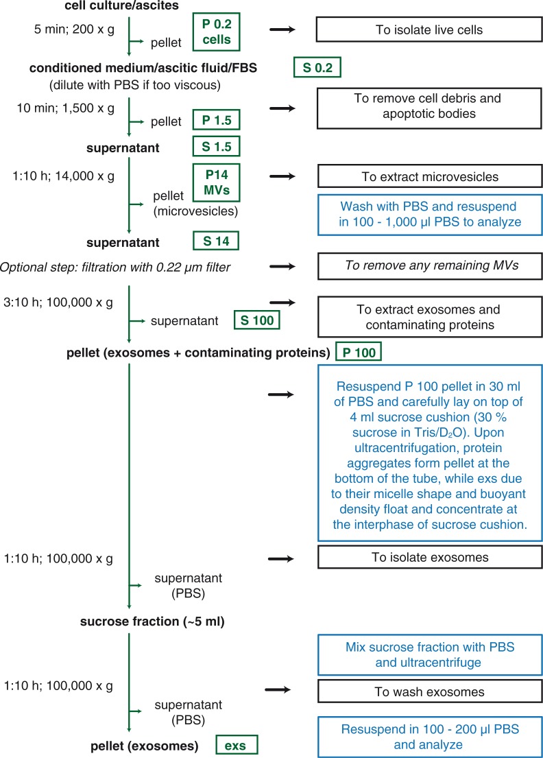
Fig. 2.
Isolation of microvesicles (MVs) and exosomes (exs) by ultracentrifugation (UC) and sucrose cushion. A schematic representation of protocol used to isolate extracellular vesicles (EVs). Black boxes describe the purpose of each centrifugation step, blue boxes contain additional instructions/explanation. Optional filtration step is indicated by italics. Green boxes depict fraction after centrifugation at relative centrifugation force (RCF) indicated by number (in thousands×g). P, pellet; S, supernatant.

