Fig. 6.
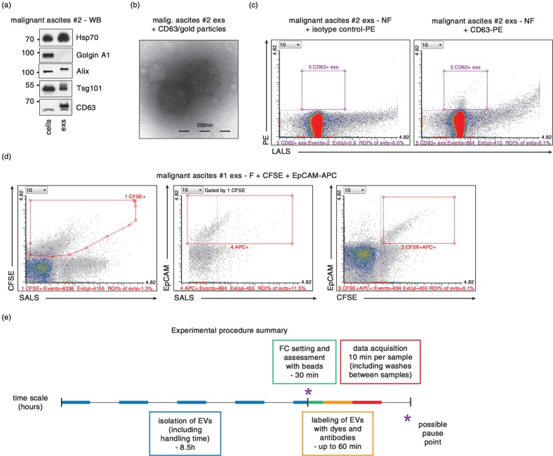
Characterization of exs derived from ascites using antibodies. (a) Presence of characteristic exs markers Alix, Hsp70 and Tsg101 as well as CD63, and absence of “antimarker” Golgin A1 (present in the cells of origin but absent from exs) was verified by western blotting (WB) in cells (P 0.2 fraction) and exs from ovarian cancer patient #2. (b) TEM of exs isolated from ascites of ovarian cancer patient #2. Exs marker CD63 is detected at the surface of exs by secondary antibodies conjugated to 10-nm gold particles. (c) CD63+, exs derived from ascites of ovarian cancer patient #2 can be detected by FC analysis of CD63-PE antibody-stained exs (right panel). Left panel, exs stained with isotype control antibody-PE. (d) Double labeling of exs from ascites of patient #1 by CFSE and EpCAM-APC antibody reveals feasibility of double labeling and demonstrates that only 11.5% of CFSE+ exs are EpCAM+. (e) Summary of timing of the presented protocol. Each step is marked in the time scale by different color. For collection of EVs from conditioned media, cells are plated a day ahead. Isolation of exs can be achieved in 1 day. Setting up the instrument and FC data acquisition can be completed within the morning of the second day. – F, 0.22 µm filtered; – NF, 0.22 µm non-filtered.

