Figure 1.
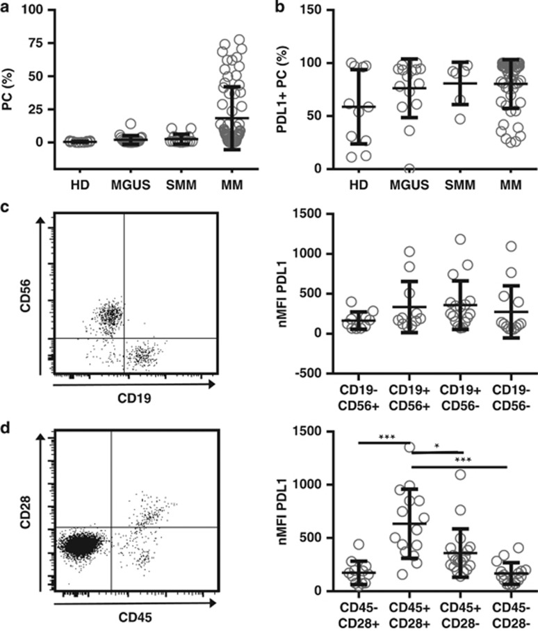
PD-L1 is expressed on malignant and normal plasma cells. (a) Bone marrow of healthy donors (HD, n=11), monoclonal gammopathy of undetermined significance (monoclonal gammopathy of undetermined significance (MGUS), n=15), smoldering myeloma (SMM, n=6), and multiple myeloma patients (MM, n=51) were analyzed by flow cytometry for percentages of plasma cells by gating on CD38+CD138+ cells after doublet exclusion. (b) Percentages of PD-L1 positivity were assessed in relation to matched isotype or FMO controls, using a mouse anti-human PD-L1 monoclonal antibody (clone 29E.2A3; BioLegend, San Diego, CA, USA). Only those samples with event numbers >100 within the PC gate were taken into account. (c) Normalized (subtraction of FMO control) median fluorescence intensity (nMFI) of PD-L1 was assessed on different PC subsets of SMM, MGUS and MM patients (n=22) according to their CD56 or CD19 expression or (d) CD28 and CD45 expression (exemplary dot blots on the left). Only those samples where clear populations were detectable and the number of events was >50 were taken into account. All data are presented as mean±s.d. Statistical significance was evaluated using analysis of variance followed by Tukey's multiple comparison test. *P<0.05, ***P<0.001.

