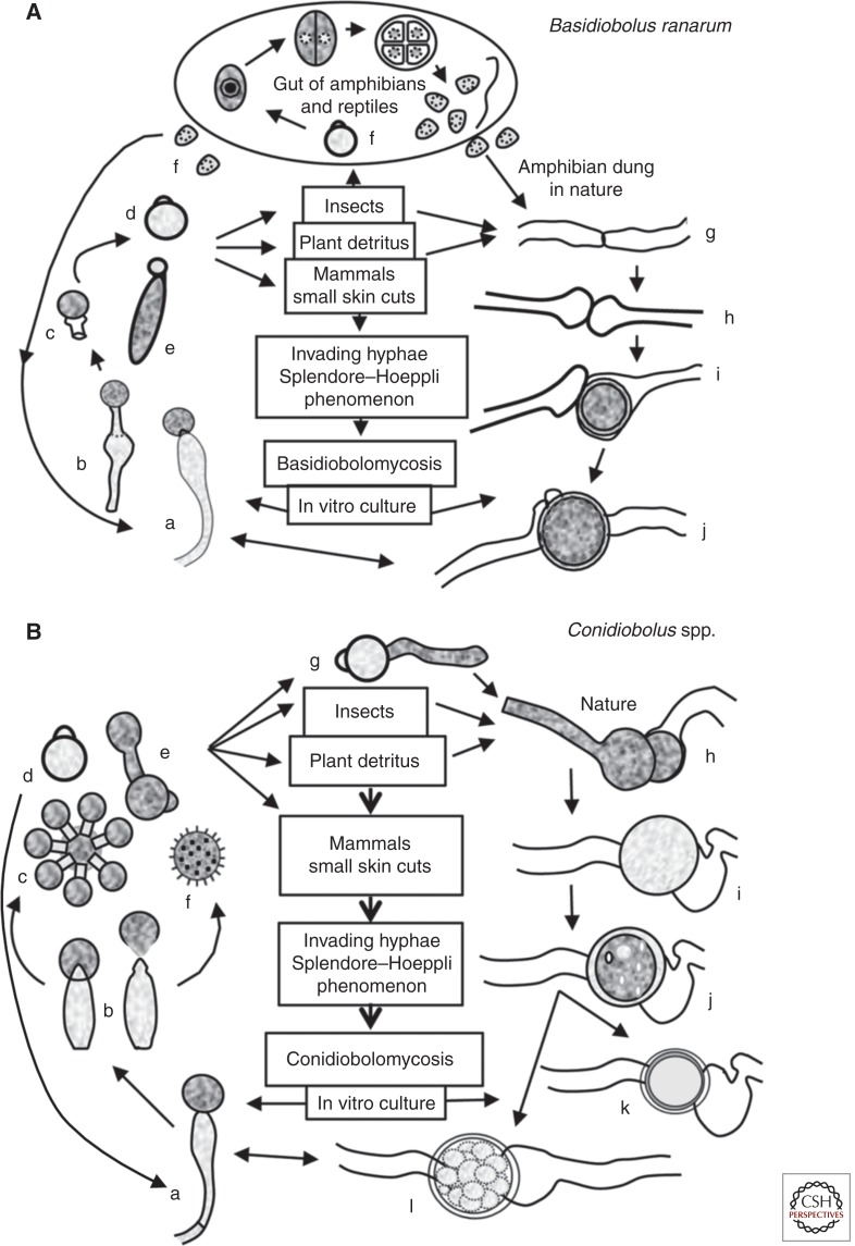
Figure 3.
The life cycles of the representative species that cause entomophthoramycosis. (A) The life cycle of Basidiobolus ranarum. The life cycle starts after a single sporangiophore (a and b) forcibly ejects a conidium (c and d) or after the formation of an elongated sticky “capilliconidium” with a terminal sticky beak (e). Insects or other animals, including mammals, could come into contact with these sticky conidia. Some reptiles and amphibians feed from insects carrying these spores, and a new cycle begins inside their gut (f). Levisohn (1927) observed that a single conidium divided and could produce >50 small spores (meristospores), which could survive in dry environments. After being released into the environment, they develop short mycelia and produce single sporangiophores; alternatively, the nuclei in the same hyphae could exchange genetic material and develope zygospores (g–j). They can also develop into structures called palmella, which resemble green algae. Mammals inhabiting areas of endemicity can contact B. ranarum propagules through trauma and develop zygomycosis (center).
(B) The life cycle of the pathogenic Conidiobolus species. A single conidiophore develops terminal conidia (a and b), which are forcibly ejected (d) using a mechanism different from that in the genus Basidiobolus (see taxonomy) and geminate (g). Most species that are pathogenic to mammals can develop replicative conidia (c and e), but the “villose” conidia (f) can only be found in C. coronatus. These conidia could develop more sporangiophores or repeat the cycle, or the nuclei of the same coenocytic hyphae could form a septum and develop into a zygospore (h–l). Drawings of k and l are examples of C. lampragues and C. incongruus zygospores, respectively.

