
Figure 1. Nuclear features of lung adenocarcinoma (hematoxylin and eosin stain; original magnification, ×400: A–E, ×600: F).
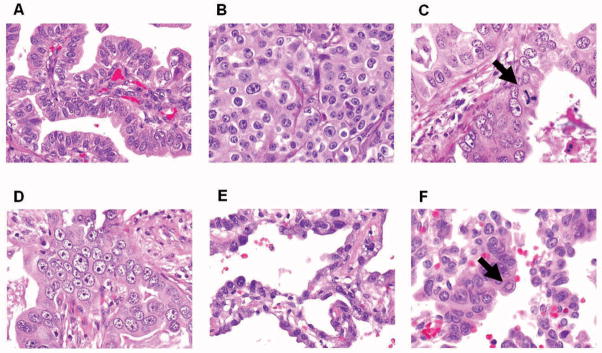
(A) Tumor cells showing mild nuclear atypia with small nuclei, intermediate nuclear/cytoplasmic (N/C) ratio, fine granular chromatin, and distinct nucleoli. (B) Tumor cells showing moderate nuclear atypia with intermediate size nuclei, and low N/C ratio. (C) Tumor cells showing large nuclei with atypical mitosis (arrow). (D) Tumor cells showing severe nuclear atypia with coarse granular chromatin, and large nucleoli. (E) Tumor cells showing high N/C ratio, homogeneous chromatin, and indistinct nucleoli. (F) Tumor cells with intranuclear inclusion (arrow).