
Figure 6.
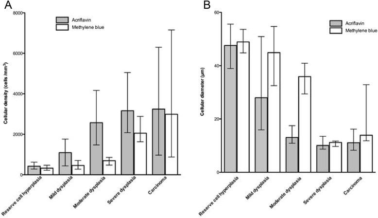
Change in cell density and diameter using FCFM according to the fluorophore (acriflavine or methylene blue) during the different stages of the malignant transformation of the hamster cheek pouch. A Median cell density (/mm2) calculated from imaging with acriflavine or methylene blue according to histological diagnosis. The data are plotted as median with range. Cell density was significantly higher in acriflavine-aided FCFM compared with methylene blue imaging. Respectively, it was 423 vs. 324 cells/mm2 in the hyperplastic stage (p = 0.006), 1093 vs. 459 cells/mm2 in the mild dysplastic stage (p = 0.006), 2574 vs. 694 cells/mm2 in the moderate dysplastic stage (p < 0.001), and 3163 vs. 2055 cells/mm2 in the severe dysplastic stage (p = 0.02). B. Median diameter (μm) measured using acriflavine or methylene blue-aided FCFM according to histological diagnosis. The data are plotted as median with range. Using acriflavine-aided FCFM, the diameter decreased during the different stages from hyperplasia to high-grade lesions (p = 0.003 between hyperplasia and mild dysplasia, p < 0.001 between mild and moderate dysplasia, p = 0.001 between moderate and severe dysplasia, p = 0.002 between moderate dysplasia and severe dysplasia/carcinoma).