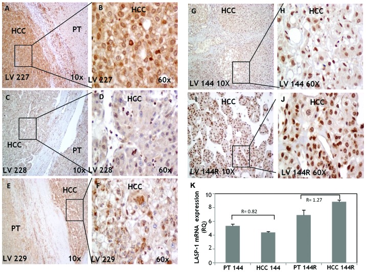
Figure 2.
The evaluation of LASP-1 protein expression in HCC and PT tissues by IHC. LV 227. Strong LASP-1 expression in HCC, weak expression of LASP-1 in adjacent non-tumors tissue (A). Higher magnification shows positive cytosolic and weak nuclear expression of LASP-1 in HCC (B). LV 228. In PT tissue LASP-1 expression was detectable only in the inflammatory cells (C); weak cytoplasmic and nuclear expression of LASP-1 in HCC (D). LV 229. Weak cytoplasmic positivity of LASP-1 in PT, moderate expression in HCC (E); strong cytoplasmic and weak nuclear staining in HCC tissue (F). LV 144. Moderate nuclear staining particularly in differentiated area of HCC tissue (G and H). LV 144 recurrence. Strong nuclear positivity in this moderate differentiated HCC, negative or very weak cytoplasmic positivity (I and J). LASP-1 mRNA expression levels evaluated by qPCR in LV144 and LV144R (K). Histograms represent RQ (relative quantification) values, bars are ± RQmax, RQmin.

