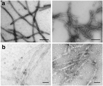
Figure 3.

Macrostructure of the AChE-PrP fibril morphotype. Negative-stained transmission electron micrographs of PrP fibrils formed in the absence (left panels) or presence (right panels) of AChE, after direct deposition onto a grid (a), or following incubation with an anti-AChE antibody and a secondary antibody conjugated to 10-nm gold nanoparticles (b). The molar ratio used was 0.5:1 (AChE:PrP). All scale bars are 100 nm.
