
Figure 2.
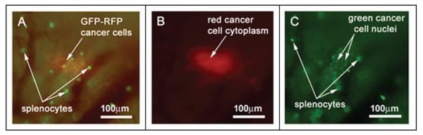
Multicolor imaging using different fluorescence emission filters allows for discrimination between cancer cells and GFP-expressing splenocytes within the native pancreas in vivo. GFP-RFP tumor-bearing animals were given a single i.v. injection of GFP-expressing splenocytes. The animals were imaged at 9 days after splenocyte injection. (A) Use of the GFP long-pass filter allows visualization of both green and red fluorescent signals from the cancer cells as well as the isolated green signal from the splenocytes. (B) The RFP narrow-band filter allows discrimination of the DsRed2 signal from the tumor cell cytoplasm. (C) The GFP narrow-band filter shows GFP signals from the cancer-cell nuclei as well as GFP-expressing splenocytes. Imaging was with the Olympus OV100.