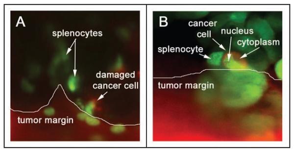
Figure 4.

IV100 Imaging of GFP-expressing splenocyte interactions with the GFP-RFP tumor margin. GFP-RFP tumor-bearing animals were given a single i.v. injection of GFP-expressing splenocytes. The animals were imaged at 9 days after splenocyte injection. The scanning laser microscope system allows high resolution imaging of single cells within the pancreatic microenvironment. (A) The GFP-RFP tumor margin with red fluorescent cytoplasm and green fluorescent nuclei are shown with adjacent green fluorescent spleen cells and damaged GFP-RFP cancer cells in the peritumoral tissue. (B) High-resolution imaging allows for observation of both cancer cell-immune cell interactions as well as individual tumor cell morphology. All images taken with IV100 20x objective.
