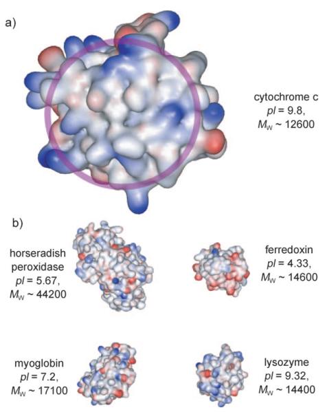
Figure 1.
Structures of a) cytochrome c (PDB ID: 1HRC)[39] and its rec ognition surface for interaction with other proteins circled in light purple and b) other proteins tested, including horseradish peroxidase (PDB ID: 1W4W),[40] ferredoxin (PDB ID: 1A7O),[41] myoglobin, (PDB ID: 1HRM),[42] and lysozyme (PDB ID: 2LYM).[43]

