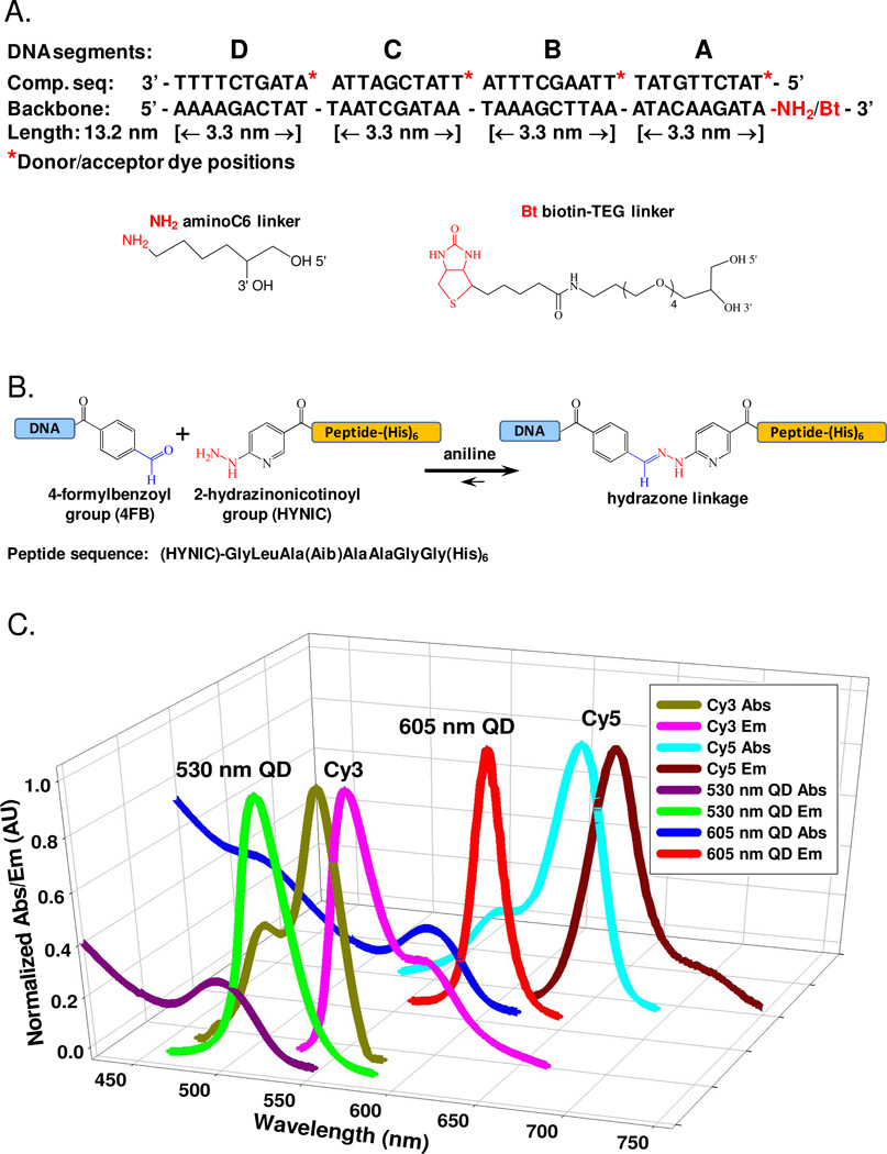
Figure 1. DNA sequences, chemoselective ligation and spectral overlap.
(A) Sequences of the DNA backbone with a 3’ amino or biotin functionalization and the complementary DNA segments (A–D) showing donor/acceptor labeling sites at the 5’ end of each. (B) Aniline catalyzed hydrazone ligation between the aldehyde (blue) of the 4FB group and the peptidyl HYNIC group (red) used to link DNA to the (His)6-peptide. (C) Plot showing the spectral overlap of the fluorophore donor-acceptor species used; Cy3-Cy5, 530 nm QD-Cy3 and 605 nm QD-Cy5.

