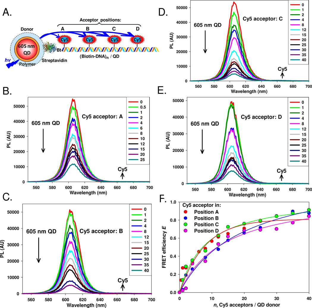
Figure 4. Construct 3: Biotin-DNA streptavidin QD assembly.
(A) Schematic of the nanoconstruct comprised of a streptavidin-functionalized 605 nm QD bound to the biotin labeled 5’ end of the DNA backbone hybridized with Cy5 acceptor DNA at positions A–D. When a position is not used, unlabeled spacer DNA is hybridized in that location. (B–E) PL spectra of 605 nm QD donors conjugated to increasing molar ratios of Cy5 labeled DNA in positions A–D, respectively. (F) Plot of FRET efficiency E for each acceptor position versus acceptor valence. Lines of best fit added.

