Figure 4.

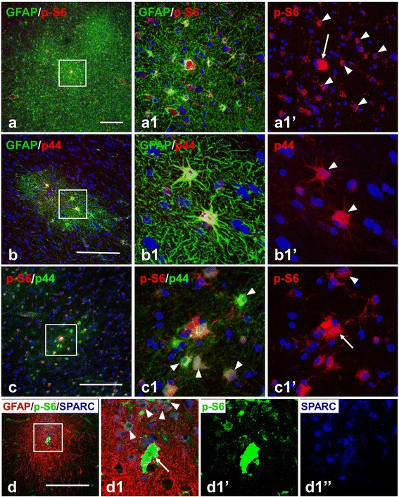
Astrocytes in microtubers reveal activation (phosphorylation) of ribosomal protein S6 (S6) and p44/42 MAPK (p44). (a) p-S6+ astrocytes (arrowheads, marked only some) in microtubers in cortical layer V. A giant p-S6+ cell is marked with an arrow. Note that several microtubers are located near each other. (b,c) p44+ astrocytes (arrowheads) in microtubers detected with polyclonal (b, red) and monoclonal (c, green) primary antibodies. Giant cell p–S6+ and p44+ is marked with an arrow. (d) Many SPARC+ fibrous astrocytes express p-S6 (arrowheads). Giant p-S6+ /SPARC+ cell is marked with an arrow. Confocal microscopy, double immunostaining, counterstaining with Nissl (a-c), triple immunostaining (d). a1, b1, c1, and d1–enlarged boxed area in a, b, c, and d, respectively. a1’, b1’, c1’,d1’ and d1”represent split a1, b1, c1, and d1 images, respectively. scale bars: 150 μm.
