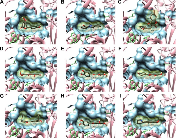
Figure 3.
Molecular surface representations of NS5B binding pocket with top docked ligands.
Notes: Conformation of top ligands (binding energy >−9 kcal/mol) inside binding pocket shown by sticks in dim gray. The protein-binding pocket is exposed in molecular surface representation (light blue), with the 12 interacting residues within 4 Å from ligand displayed by green sticks. Docking view of naringenin (A), tryphanthrine (B), dicoumarin (C), swertianin (D), diosmetin (E), apigenin (F), honokiol (G), luteolin (H), and thaliporphine (I).
Abbreviation: NS5B, nonstructural protein 5B.

