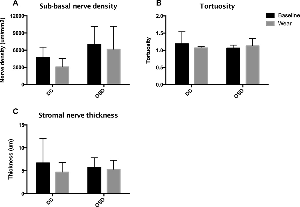
Figure 3.
Corneal nerve structure and morphology assessed by sub-basal nerve density, tortuosity and stromal nerve thickness. Measurements at baseline and after long-term device wear were compared and categorized by patient diagnosis. There was no change in sub-basal nerve density, sub-basal tortuosity, or stromal nerve thickness over time for either diagnosis group, but DC patients showed a lower density of sub-basal nerves compared to OSD patients.

