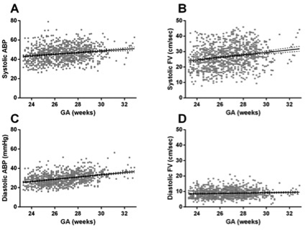
Figure 2.
ABP and CBF velocity are shown as a function of GA. A and B) Systolic ABP and CBF velocity both trend upward between 23 and 33 weeks gestation (r = 0.26 and 0.24 respectively, p<0.001). C and D) While diastolic ABP trends upward by more than 1 mm Hg/week gestation in this same developmental period, diastolic CBF velocity shows very little increase, clustering near the functional 0 reading of the transcranial Doppler used (r = 0.43 and 0.009; p < 0.001 and p = 0.003 respectively).

