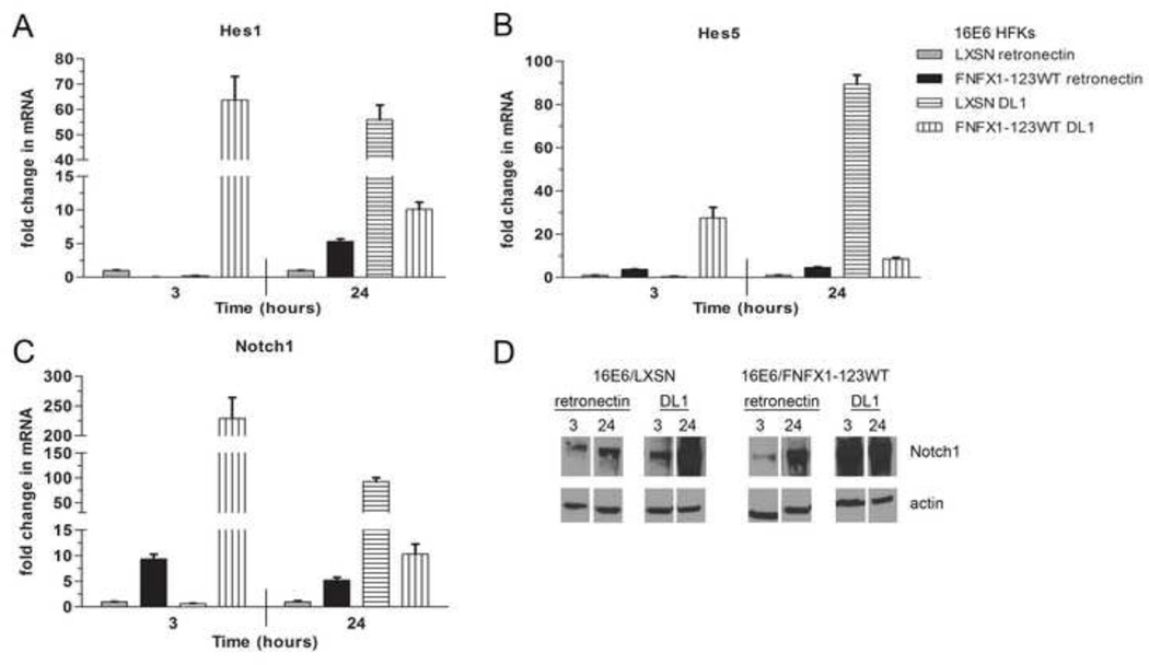
Figure 3. DL1 activated Notch1 canonical pathway signaling.
16E6/LXSN and 16E6/FNFX1-123WT HFKs were plated on dishes coated with purified Delta-1 ligand (DL1) immobilized on retronectin or retronectin alone. Whole cell extracts were harvested for RNA and protein at the indicated times. Similar results were found in at least three independent experiments. (A) Relative levels of Hes1 mRNA (B) Hes5 mRNA and (C) Notch1 mRNA were calculated using the ΔΔCT method, normalizing mRNA levels to GAPDH within each sample. Values shown were the mean fold change in each sample compared to the 16E6/LXSN vector control plated on retronectin. Error bars represent the standard deviation for triplicate samples. (D) Representative immunoblot of Notch1 protein in whole cell extracts of 16E6/LXSN and 16E6/FNFX1-123WT HFKs at the indicated times. Actin shown as a loading control.

