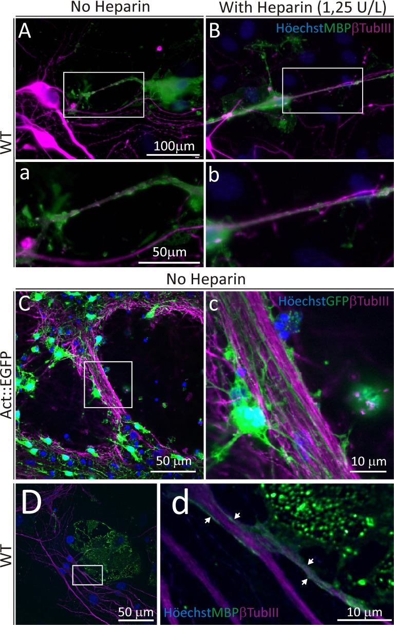
Fig 7. Confocal and epi-fluorescence microscopy images of WT neuron and Act::EGF NS co-cultures.
Axonal fibers are labelled with βTubulin III (magenta) for all images. A, B) Epi-fluorescence images of MBP+ (green) and βTubIII cells belonging to bFGF/PDGF-BB cultures treated without (A) or with Heparin (B) during proliferation. The insets in A and B are shown in a and b, respectively. C, D) Confocal images of co-cultures of neurons and bFGF/PDGF-BB (with no added Heparin) pre-treated neurospheres. C) Image of EGFP+ cells from Act::EGFP mice and β-TubIII+ neurons. D) Confocal image of an MBP+ OL (from WT-derived neurospheres) interacting with a βTubIII+ neuron projection. The insets C and D is shown enlarged in c and d, respectively. Scale bar in A = 100 μm for A and B, scale bar in a = 50 μm for a and b.

