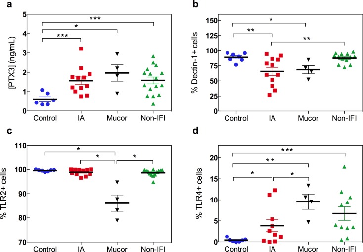
Fig 1. Pattern recognition receptors in hematological patients with IMI.
(a) Serum levels of PTX3 (ng/mL) measured by immunoassay in healthy controls (blue circles; n = 6), hematological patients with IA (red squares; n = 12) or mucormycosis (black inverted triangles; n = 4) and non-IFI hematological controls (green triangles; n = 16) are shown. *p<0.05, **p<0.01, and ***p<0.005 using the unpaired two-tailed Student’s t-test. (b-d) Surface expression of dectin-1 (b), TLR2 (c) and TLR4 (d) on monocytes was measured by flow cytometry. Gating on CD45highCD14+ cells was performed in order to avoid interference of the analysis by potential blasts in leukemic patients with residual disease. Dot plots represent the percentage of monocytes (CD45highCD14+ cells) expressing dectin-1, TLR2 or TLR4 in peripheral blood samples from healthy controls (blue circles; n = 7), hematological patients with IA (red squares; n = 12) or mucormycosis (black triangles; n = 4) and non-IFI hematological controls (green triangles; n = 13). *p<0.05, **p<0.01 using the unpaired two-tailed Student’s t-test. All data are shown as mean ± s.e.m.

