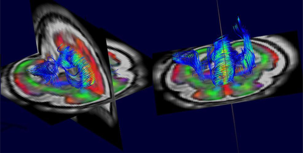
Figure 3.
An example of between slice motion corrected high resolution 3D diffusion tensor imaging from combined multi-slice and multi-plane DWI. This allows tractography based analysis to quantify the development of white matter connections within the fetal brain based on the estimates of water diffusion over the brain. Note also the color coded direction map of the surrounding anatomy that highlights the highly radial structure of the cortex at this early stage of development. This contrasts with the cortical microstructure of the adult cortex which provides no directional constraints on the diffusion of water..

