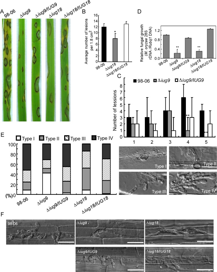
Fig 8. IUG9 and IUG18 are involved in pathogenicity of M. oryzae.
(A) Disease symptoms were reduced on rice leaves inoculated with Δiug9 and Δiug18 mutants. Conidial suspension (5 x 10⁴ spores/ml) of the wild-type strain 98–06, mutants and complemented strains were inoculated on rice (cv. LTH), and incubated for 7 days. (B) Bar chart of mean lesion density of seedlings infected with isolate 98–06 and the Δiug18 mutant per unit area. Mean lesion density was significantly reduced in Δiug18 mutant infections. Error bars represent the standard deviation and asterisks represent significant differences (P<0.01). (C) Quantification of lesion types (0, no lesion; 1, pinhead-sized brown specks; 2, 1.5-mm brown spots; 3, 2–3-mm grey spots with brown margins; 4, many elliptical grey spots longer than 3 mm; 5, coalesced lesions infecting 50% or more of the leaf area) reveals no difference in lesion types 1–3; however, the Δiug9 mutant make rarely lesions of types 4 and 5. Lesions were photographed and measured or scored at 7 days post-inoculation (dpi) and experiments were repeated twice with similar results. (D) Severity of blast disease was evaluated by quantifying M. oryzae genomic 28S rDNA relative to rice genomic Rubq1 DNA (7 days post-inoculation). Mean values of three determinations with standard deviations are shown. The asterisks indicate a significant difference from the 98–06 (P < 0.01). (E) Percentage of difference infection hyphae type (I = no infection hyphae; II = only one infection hyphae; III = two or three branches of the infection hyphae; IV = more than three branches of infection hyphae), occupied by each strain in the reverse side cells of barley 32 h after inoculation. The total number of appressorium-mediated penetration and infection is indicated (top right corner, N = 100). (F) Typical infection sites of rice leaf sheath inoculated with 98–06 strain, Δiug18, Δiug9 mutants, and complemented strains, showing greater fungal proliferation and tissue invasion by the wild-type strain. Infectious growth was observed at 30 hpi. Bars = 50 μm.

