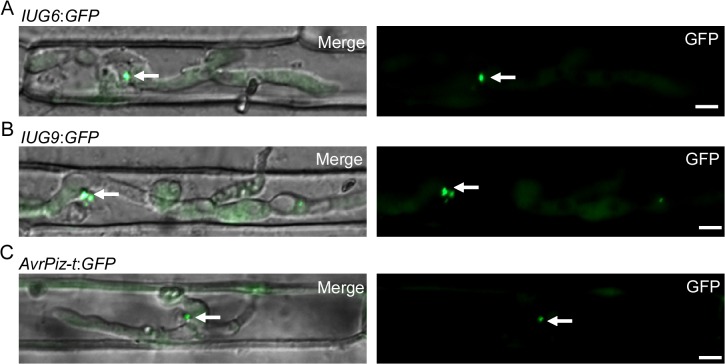
Fig 9. Iug6 and Iug9 proteins accumulate at BICs in sheath epidermal cells.
(A) Cellular localization of Iug6:GFP in M. oryzae during biotrophic growth on epidermal rice cells at 27 hpi. Fluorescence was observed accumulating preferentially at BICs. Merged DIC and GFP images and GFP fluorescence alone are shown. BICs are indicated by arrows. Bars = 10 μm. (B) Secretion of Iug9:GFP at 27 hpi. Fluorescence was at BICs. Bars = 10 μm. (C) AvrPiz-t:GFP was observed preferential BIC localization at 30 hpi. Bars = 10 μm.

