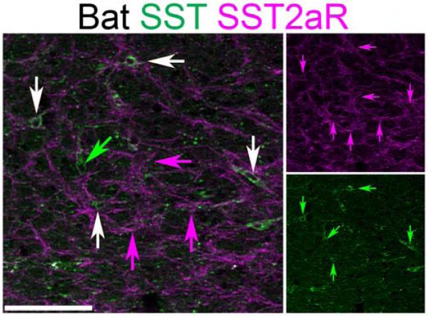
Figure 5.
Bat ventrolateral medulla SST neurons co-express SST2aR. Two-color confocal projection images showing partial co-expression of SST (green) and SST2aR (magenta) within the approximate region of the bat preBötC. Single-color images are shown to right. White arrows indicate SST and SST2aR co-expression. Green arrow indicates SST neuron lacking SST2aR expression. Magenta arrows indicate SST2aR neurons lacking SST expression. Scale bar, 100 µm.

