
Figure 3.
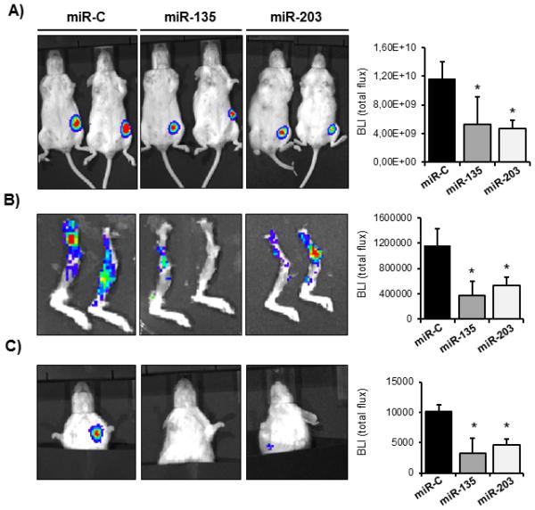
miR-135 and miR-203 impair tumor growth and spontaneous metastasis to bone and lung. A, tumor growth in the mammary fat pad in vivo was determined by quantifying the bioluminescence signal after transplanting MDA-MB-231-luc cells ectopically expressing miR-135 and miR-203, followed by intratumoral injection of synthetic miRNAs. Tumor growth was quantified by the bioluminescence signal using the LivingImage software. B, spontaneous metastasis from the orthotopic site to bones and C, to the lung was visualized by bioluminescence. Ectopic expression of miR-135 and miR-203 significantly impaired spontaneous metastasis. Bioluminescence intensity was quantified using the LivingImage software and data are represented as means ± SD of each group. *significant difference (p< 0.05) between indicated miRNA and miR-C, Tukey’s post-hoc test. Representative mice from n= 6-12 in each group are shown.