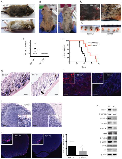
Figure 1. Loss of PDK1 inhibits the onset of melanoma development and delays metastasis.
(A–B) Representative images of PDK1 WT (BrafV600E::Cdkn2a−/−::Pten+/+::Pdk1+/+) or PDK1 KO (BrafV600E::Cdkn2a−/−::Pten+/+::Pdk1−/−) mice 21 days after administration of 4-HT. (C–D) Representative images of the dorsal skin (C) and lymph nodes (D) from PDK1 WT and PDK1 KO mice 38 days after systemic administration of 4-HT. (E) Quantification of tumors in PDK1 WT and PDK1 KO mice (N = 12 and 8, respectively). (F) Kaplan-Meier survival curves of PDK1 WT and PDK1 KO mice (N = 16 and 18, respectively). P < 0.001 by log-rank (Mantel-Cox) test. (G–H) H&E stained (G) and S100 immunostained (H) skin sections from PDK1 WT and PDK1 KO mice 38 days after 4-HT administration. Bars = 100 μm. (I–J) H&E stained (I) and S100 (J) immunostained lymph nodes from PDK1 WT and PDK1 KO mice 38 days after administration of 4-HT. Bars = 100 μm. Graph shows mean ± SEM of S100+ cells from 3 animals per genotype. *P < 0.005. (K) Western blot analysis of the indicated proteins in primary melanoma cultures derived from PDK1 WT and PDK1 KO mice.

