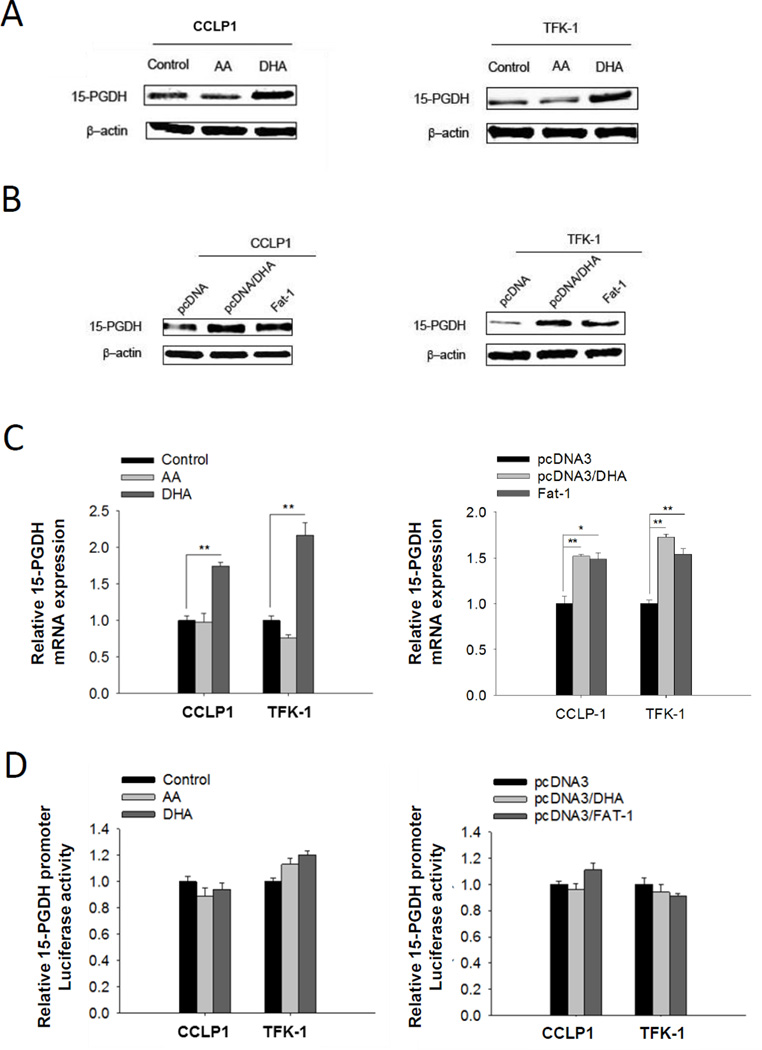
Figure 1. ω-3 PUFAs induces 15-PGDH expression in human cholangiocarcinoma cells.
(A) 15-PGDH protein level increased in cholangiocarcinoma cells cultured with ω-3 PUFA DHA but not with ω-6 PUFA AA. CCLP1 or TFK-1 cells were synchronized by serum deprivation, and then maintained in serum-free medium containing 50 µM DHA or AA for 12 h; (B) 15-PGDH protein level increased in cholangiocarcinoma cells expressing Fat-1 which converts ω-6 PUFA to ω-3 PUFA. CCLP1 or TFK-1 cells stably transfected with Fat-1 expression vector or control vector were synchronization by serum deprivation. The cells weer treated with or without 50 µM DHA in serum-free medium for 12 h. Total protein was analyzed by Western blotting with 15-PGDH antibody. β-actin was measured as a reference gene. (C) DHA treatment or Fat-1 gene expression increases 15-PGDH mRNA level in cholangiocarcinoma cells. 15-PGDH mRNA was measured by real-time PCR. Results were normalized to control group; the data were shown with mean±SE. *P<0.05, **P<0.01; (D) 15-PGDH promoter activity was not influenced by DHA treatment or Fat-1 expression in cholangiocarcinoma cells. CCLP1 or TFK-1 cells were co-transfected with 15-PGDH promoter-luc3 and pRL-TK plasmid which code Renilla luciferase. 72h after transfection, Luciferase activity was measured. Results were normalized to control group. Data were shown with mean±SE.

