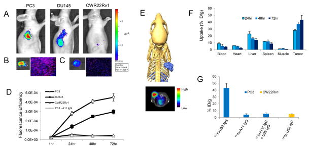
Figure 4.
Molecular imaging and biodistribution of U33 IgG in prostate cancer xenografts. (A) Near-infrared (NIR) optical imaging of prostate cancer xenografts using AF680-U33 IgG. Mice bearing PC3, DU145 or CWR22Rv1 xenografts were tail-vein injected with 2 nmol of AF680-U33 IgG and imaged using NIR optical imaging. The images shown are representative of n=3 mice/xenograft and were acquired 72hrs post-injection. (B) The resected PC3 tumor at 72hrs fluoresence intensity (left) and a tumor section demonstrating probe penetration and localization by fluoresence microscopy (right). (C) Probe fluoresence intensity (left) and localization (right) in the liver of a PC3 xenograft mouse. (D) Graph depicting the localization of AF680-U33 IgG as fluoresence efficiency of the tumor ROIs for the mice imaged using NIR optical imaging. Included in the graph are the data for the mice imaged with the isotype control AF680-A11 IgG in PC3 xenografts. (E) SPECT imaging with 111In-U33 IgG in a PC3 xenograft model. Depicted are SPECT/CT images shown as a three-dimensional volume rendering of the SPECT data (blue) overlaid onto surface rendered CT data and a reconstructed transverse view using a rainbow color scale to show uptake (below). Image is representative of n =3 mice imaged with 111In-U33 IgG at 72hrs post-injection. Each animal for imaging received 2.5μg of antibody corresponding to 220 μCi of activity. (F) Probe biodistribution was determined by radioactivity assays in PC3 tumor bearing mice (n = 4 for each time point). Tissues were harvested at 24, 48 and 72hrs after injection of 111In-U33 IgG (25 μCi). Probe uptake is reported as percent injected dose per gram (%ID/g). (G) Tumor uptake specificity measured at 72hrs post-injection (n = 4 mice for each treatment). PC3 xenograft bearing mice were treated with isotype control 111In-A11 IgG (25μCi) and 111In-U33 IgG blocked (80% reduction) by i.v. pre-injection of 200 μg of cold U33 IgG. Probe uptake in CWR22Rv1 xenografts is also depicted.

