


Figure 2. The genetic organization of the pCW3 tcp locus.
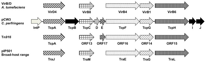
Proteins encoded by the various genes are stated below the arrows. The patterned arrows indicate Tcp proteins involved in pCW3 conjugative transfer, black arrows depict non-essential Tcp proteins and the grey arrow the IntP protein, which is currently uncharacterized. Proteins with sequence, functional or structural similarity from the paradigm VirB/D system from the Ti plasmid from the Gram-negative Agrobacterium tumefaciciens, the conjugation region from Tn916 and the broad-host range plasmid pIP501 from the Gram-positive Streptococcus agalactiae are represented by arrows with similar patterns. Each of these conjugation regions has homologs of the putative coupling proteins, VirD4 and TcpA, VirB8-like proteins, VirB4-like ATPases, VirB1-like lytic transglycolases and VirB6-like proteins. Based on data from [128, 129, 135].