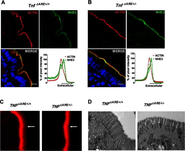
Fig. 4.
NHE3 localization and microvillar length in the distal ileum of Tnf ΔARE+/+ and +/− mice. a In the distal ileum of Tnf ΔARE+/+ mice, NHE3 was found both in the intervillar cleft/terminal web region (the NHE3 which colocalizes with the peak of the F-actin signal) and in the microvillar region (the peak that is more extracellular to the peak of the F-actin signal). b In the BBM of Tnf ΔARE+/− mice, overall NHE3 immunofluorescence was not decreased compared with noninflamed littermates, but the peak of NHE3 was closer to that of the F-actin signal compared to the WT controls. Images are representative of three individual experiments done in at least three pairs of mice. Scale bar represents 10 μM. c High magnification of F-actin staining in the BBM revealed that the microvillar staining (hazy red zone pointed to with white arrowhead) was decreased in the distal ileum Tnf ΔARE+/− compared to Tnf ΔARE+/+. Figure represents data observed in three mice pairs. d Electron microscopy pictures taken from Tnf ΔARE+/+ and Tnf ΔARE+/− mice distal ileum showing reduced microvilli lengths in the Tnf ΔARE+/− mice; data represents the results from two pairs of mice. Scale bar represents 0.5 μM

