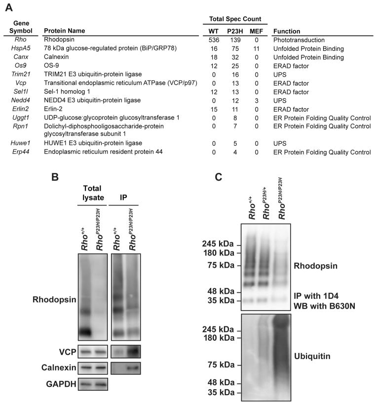
Figure 7.
P23H rhodopsin protein co-immunoprecipitates with ERAD components and is strongly ubiquitinated in photoreceptor cells. (A) ERAD-related proteins that co-immunoprecipitated with P23H rhodopsin were identified by mass spectrometry. Total spectra counts of the proteins co-immunoprecipitated with WT or P23H rhodopsin from three experimental mass spectrometry analyses are shown. Immunoprecipitation of MEF cell samples served as experimental control. (B) Rhodopsin was immunoprecipitated from retinal lysates of Rho+/+ and RhoP23H/P23H mice at P15. The elutions were concentrated by chloroform/methanol precipitation. Rhodopsin, VCP, and calnexin proteins were detected by immunoblotting. (C) Rhodopsin was immunoprecipitated from retinas of Rho+/+, RhoP23H/+ and RhoP23H/P23H mice at P15, and rhodopsin (top panel) and ubiquitin (bottom panel) levels were detected by immunoblotting.

