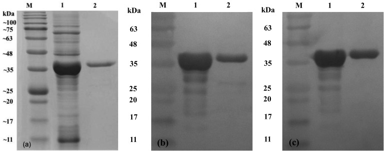
Fig. 1.
Immunoblotting of the rOmpH used in this study. The samples were run on a 12.5% SDS-polyacrylamide gel (a) and transferred to a nitrocellulose membrane. Immunoblotting of the rOmpH was done by probing with chicken sera against the rOmpH (b) or the anti-HisG-HRP antibody (c). Lane M, protein ladder; lane 1, cell lysates of the E. coli host (prior to purification); and lane 2, rOmpH fraction purified from the electroelutor. The numbers on the left indicate the positions of the molecular mass standards (in kilodaltons).

