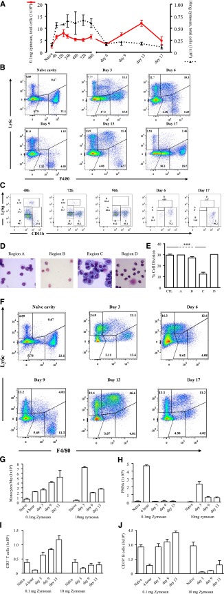
Figure 1.
Inflammation in response to low- vs high-dose zymosan in the mouse peritoneum. (A) Either 0.1 or 10 mg of zymosan was injected into the peritoneal cavity of separate groups of mice. (B) Polychromatic flow cytometry is shown carried out on inflammation driven by 0.1 mg of zymosan, highlighting monocyte/macrophage populations, whereas (C) depicts, among other cells types MDSCs, alongside their (D) histological appearance and ability to (E) suppress T-cell proliferation. (F) In contrast, flow cytometry is shown carried out on inflammation driven by a more aggressive dose of 10 mg of zymosan, highlighting monocyte/macrophage populations, whereas (G-H) summarizes the relative temporal profiles of monocyte/macrophages andPMNs in these 2 models. (I-J) Profiles of lymphocytes are shown. Data are presented as mean ± SEM for n = 8 mice/group. ***P < .005.

