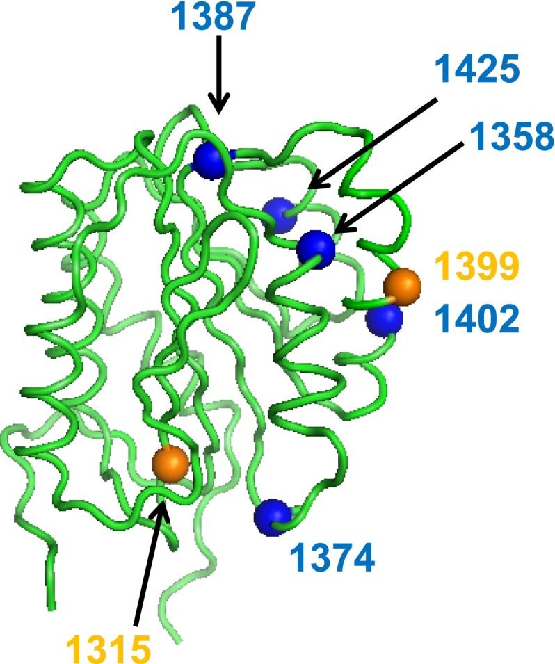
Figure 1.
Location of VWF A1 domain sequence variations and their effect on collagen 4 binding. The VWF A1 domain crystal structure30 is shown here with orange spheres to indicate Zimmerman Program A1 domain sequence variations found in type 1 subjects, and the blue spheres indicate sequence variations found in type 2M subjects that were associated with reduced or absent VWF–collagen 4 binding.

