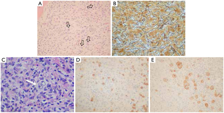
Figure 1.
(A) Diffuse involvement of bone marrow by HL with the identification of Reed-Sternberg cells (arrows) in an appropriate background (H&E, ×10); (B) Diffuse bone marrow fibrosis observed with methenamine-silver stain (×100); (C) Focal pattern of BM involvement in HL with the presence of a large binucleated Reed-Sternberg cell (arrow) (H&E, ×100); (D) Immunohistochemistry show some CD15+ Reed-Sternberg cells (IHC,×40); (E) Immunohistochemistry with expression of CD30 antigen in an adequate cellular background by Reed-Sternberg cells (IHC, ×100).

