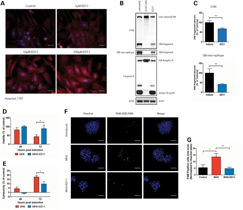
Figure 2.
ED11 penetrates the cell membrane, inhibits caspase-6 activity intracellularly and protects cells from mHtt-induced toxicity. (A) Representative images of MEF cells incubated with the indicated concentrations of ED11 and labeled with anti-TAT antibody. (B) Western blots of HEK293 cells co-transfected with caspase-6 and Htt, and stained with mab2166 and 586 neo-epitope antibodies. (C) Quantification of the Htt fragment generated by caspase-6 that was detected by mab2166 and 586 neo-epitope antibodies (n = 4–8). (D) Viability measurement of 145Q-mHtt-expressing, serum-deprived PC12 cells by Alamar blue viability assay (n = 5). (E) Cell death measurement of 145Q-mHtt-expressing, serum-deprived PC12 cells by LDH release assay (n = 5). (F) Caspase activity measurement of 145Q-mHtt-expressing, serum-deprived PC12 cells by incubation with FAM-VEID-FMK. (G) Quantification of caspase activity-dependent FAM-VEID-FMK fluorescence signal normalized to Hoechst nuclear staining (n = 7). Scale bars indicate 50 µM. *P < 0.05, **P < 0.01 one-way ANOVA followed by Tukey's post hoc test. All data are expressed as mean ± SEM.

