
Figure 4.
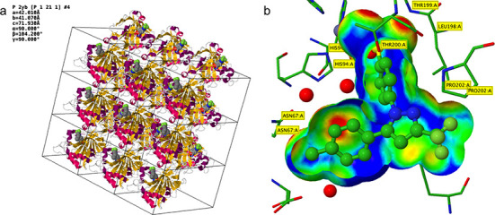
Two new custom rendering options on the Jmol /JSmol page demonstrated using PDB ID 1OQ5 (Celecoxib bound to Carbonic Anhydrase II) (41). (a) Rendering of the crystal packing (3 × 3 unit cells in the ab plane), carbonic anhydrase II (cartoon style) and Celecoxib (CPK style). (b) Jmol rendering of the Celecoxib (ball-and-stick style) in carbonic anhydrase II binding site (stick style). The trimmed ligand VDW surface is color-coded by the distance of the surface to the nearest side chain atom (short contact distances, red; optimal hydrophobic interaction distances, green; longer distances, blue). The red regions (close contacts) at the top of the surface represent hydrogen bonds and metal coordination of the sulfonamide group of Celecoxib with THR 199 and Zinc.