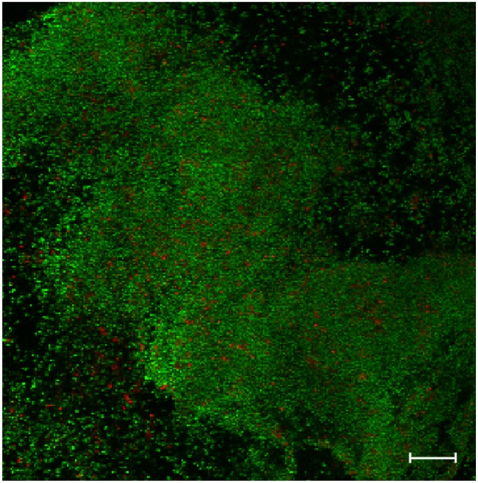
Figure 1.
Confocal laser scanning micrograph of a 24 h biofilm. The biofilm of Staphylococcus epidermidis RP62A was grown in vitro on hydrophilic acrylic intraocular lens, and was visualized after staining using the live/dead viability stain, which contains SYTO9 (green fluorescence, live cells) and propidium iodine (red fluorescence, bacterial cells that have a defective cell membrane, which is indicative of dead cells). Magnification X 400, scale bar is 20 µm.

