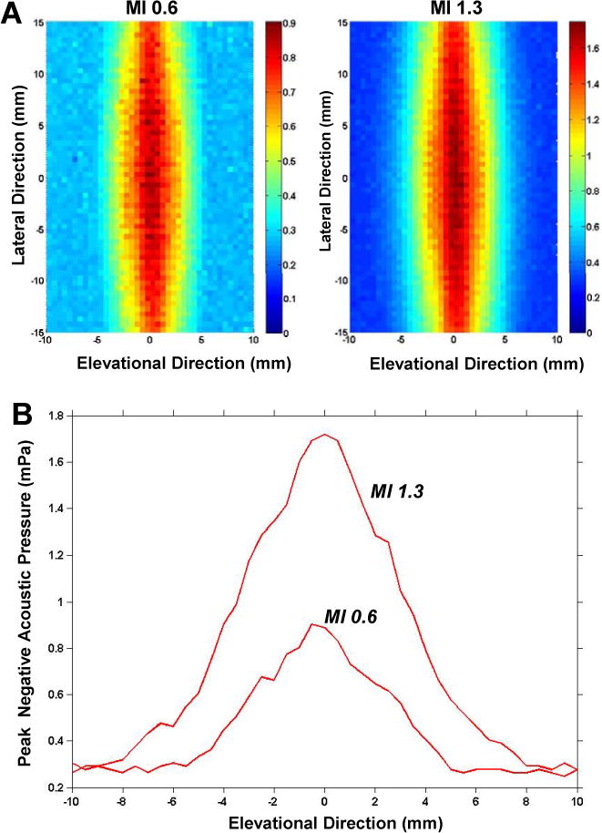
Figure 1.

(A) Peak negative acoustic pressure measurements in the lateral and elevational dimension for the two different therapeutic ultrasound conditions. Pressure measurements are color-coded according to the scale denoted in units of MPa. (B) Graphic representation of the peak negative acoustic pressure in the elevational dimension at the center of the probe, correlating to the length of muscle exposed when imaging in the short-axis plane.
