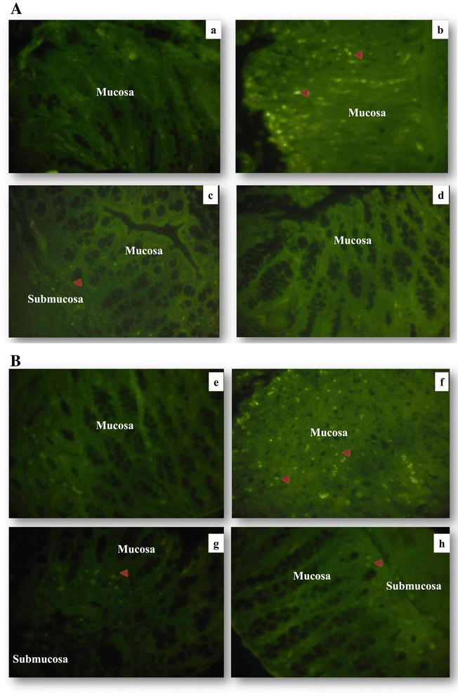
Figure 7.

Representative images of immunofluorescence staining. Distal colon tissues from mice were stained with anti-iNOS (a–d) and anti p65 NF-κB subunit (e–h). Colonic tissue stained with anti-iNOS (a) CTRL group, (b) DSS group, (c) LL/DSS group, (d) LL group. Colonic tissue stained with anti-p65 subunit NF-κB (e) CTRL group, (f) DSS group, (g) LL/DSS group, (h) LL group. Red arrow indicates iNOS or NF-κB staining, all magnifications are × 40.
