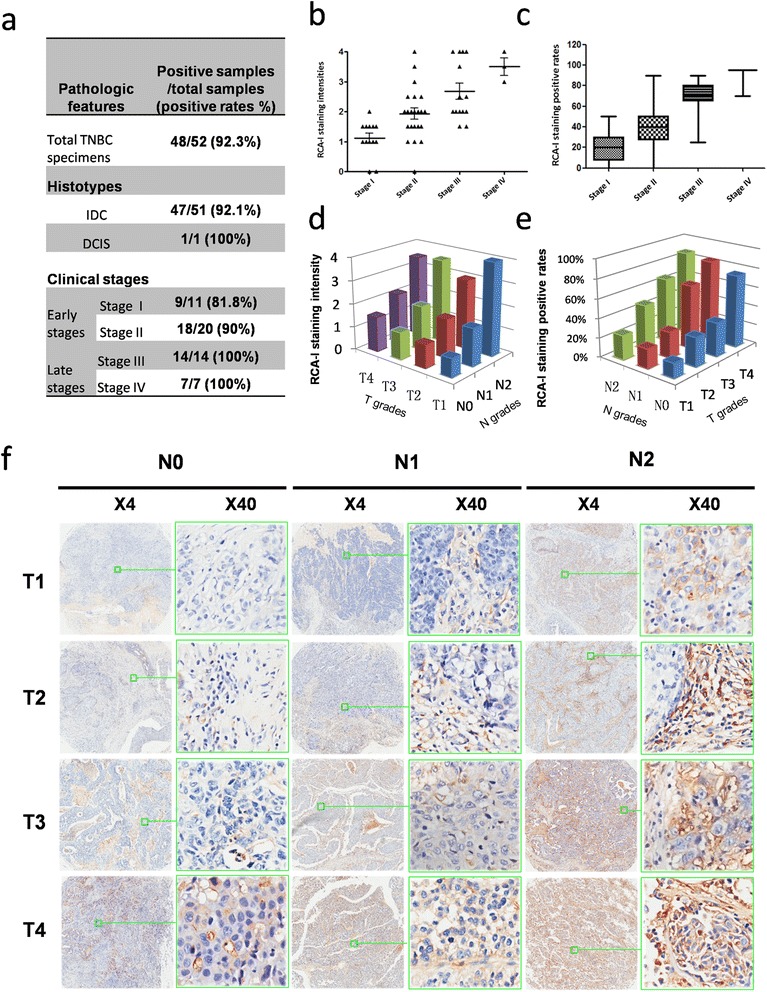
Figure 3.

Evaluation of RCA-I binding to clinical TNBC samples in a tissue microarray. (a) Overall, a very high proportion of these samples was bound by RCA-I. There was also a dependence of the rate of positive RCA-I labeling on the clinical stage of the tissue, with later-stage tissues exhibiting a higher binding rate than early-stage tissues. (b, c) The dependence of the mean intensities and positive rates of RCA-I membrane staining on the tumor stages. (d, e) The dependence of the mean intensities and positive rates of RCA-I staining on the T and N grades. (f) Representative views of TNBC tissues of different TNM grades that were incubated with biotinylated RCA-I and Cy3-conjugated streptavidin. RCA-I, Ricinus communis agglutinin I; TNBC, triple-negative breast cancer.
