
Figure 3.
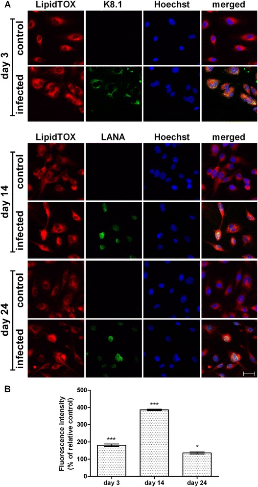
Neutral lipid detection and quantification in HHV8-infected HUVEC cells by LipidTOX dye. HUVEC cells were infected with HHV8 as described in Figure 1. 24 h before the indicated times, cells were seeded at a density of 2.0 × 10⁵ in 35 mm glass-bottomed dishes and cultured at 37°C in a 5% CO2 incubator in a growth medium. On days 3, 14 and 24 post infection, cells were fixed and treble-stained with firstly HCS LipidTOX™ Red Neutral Lipid stain (red), followed by an immunocytofluorescence method for identifying lytic (K8.1) and latent (LANA) viral-antigens with FITC-conjugated secondary antibodies (green) in the same cells counterstained with Hoechst 33258 for nuclei (blue), for details see Methods (panel A). The bar in the figure is 30 μm. Panel B represents the quantitative analysis of HCS LipidTOX™ Red Neutral Lipid stain fluorescence intensity. This method allowed the quantification of the neutral lipids in LDs in HHV8-infected cells alone. At least 10 microscopic fields were individually analysed for each experimental group. Normalized data represent the percentage of the mean density value (intensity per pixel) ± SE. Significance was set up when p < 0.05 (*) or p < 0.001 (***) vs. respective control (ANOVA and Fischer’s LSD as post hoc test).