
Figure 4.
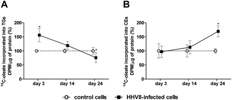
TG and CE synthesis in HHV8-infected and control HUVEC cells. HUVEC cells were infected with HHV8 as described in Figure 1. On days 3, 14 and 24 post infection, 1.0 × 10⁶ cells were incubated for 4 h in a medium containing [14C]-oleate bound to bovine serum albumin (BSA). Subsequently, cells were washed with ice-cold PBS and lipids extracted with acetone. Neutral lipids were separated by thin layer chromatography (TLC), and the incorporation of [14C]-oleate into TGs (panel A) and CEs (panel B) was determined as described in Methods. Data were reported as mean ± SE. Significance was set up when p < 0.05 (*) vs. respective control (t-test).