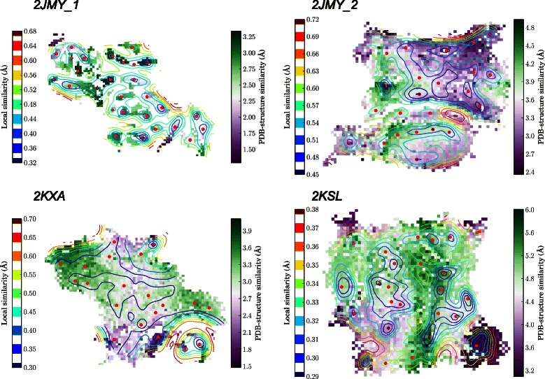
Figure 6.

Clustering of the conformations obtained by the i BP algorithm. Self-organizing maps describing the clustering of the conformations obtained by the iBP algorithm on 2JMY, 2KXA and 2KSL. The contour plots (lines) represent the local similarity between the clustered conformations. The color scales (on plot left) extend from blue to red (from very similar to very dissimilar conformations). The small red points are drawn on the SOM neuron for which the largest local similarity is observed between conformations. Each SOM neuron is colored according to the average value of the coordinates RMSD of the neuron conformations with respect to the PDB structure. The color scales extend (on plot right) from purple to green (from very similar to very dissimilar to the PDB structure). The similarity between SOM neurons as well as the RMSD to the PDB structure are expressed in Å for comparison purposes.
