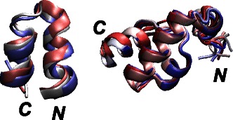
Figure 7.

Superimposed 2KXA and 2KSL conformations. Superimposition of 2KXA and 2KSL conformations extracted from the SOM, as the ones displaying the minimum coordinates RMSD with respect to the first conformer of the corresponding PDB structures. The N and C terminal extremities are labeled, and the conformations, drawn in cartoon, are colored from blue to red, according to the conformational index.
